Recombinant Antibodies

Introduction

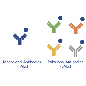
Recombinant antibodies (rAbs) are monoclonal antibodies that are generated in vitro using synthetic genes. Unlike monoclonal antibodies (mAbs) that are produced using traditional hybridoma-based technologies, rAbs do not require hybridomas or animals in the production process.

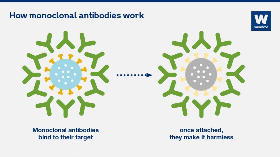
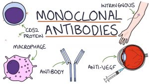
Both monoclonal and recombinant antibodies can be used in biomedical science and toxicology research, and are effective therapeutics for cancer, autoimmune disorders, and many other diseases.

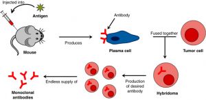
However, while monoclonal antibodies have become one of the most common tools in biomedical science and medicine due to their ability to bind to and neutralize or destroy cell-specific antigens, the ascites method of production causes significant pain and discomfort. animals used in the process.

As such, the governments of Australia, Germany, the Netherlands, and the United Kingdom banned it in favour of in vitro methods. The United States also supports the use of in vitro methods as the default procedure for the production of mAbs.

However, it is important to note that in vitro methods involving hybridomas also have their own limitations, including the following:

  • Require the immunization and subsequent euthanasia of the animals used in the process.
  • Slow and laborious.
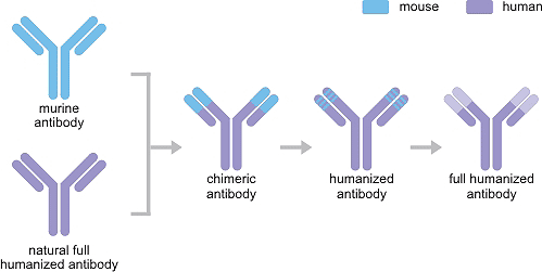
  • They often cause immune reactions, so antibodies need to be altered and “humanized” before they can be given to patients.

Recombinant antibody production

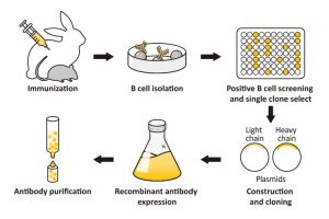
Basically, recombinant antibodies are monoclonal antibodies generated in vitro using synthetic genes. The technology involves retrieving antibody genes from source cells, amplifying and cloning the genes into an appropriate phage vector, introducing the vector into a host (bacteria, yeast, or mammalian cell lines), and achieving the expression of adequate amounts of functional antibodies.

Recombinant antibodies can be cloned from any species of the antibody-producing animal provided that appropriate oligonucleotide primers or hybridization probes are available. The ability to manipulate antibody genes also makes it possible to generate new antibodies and antibody fragments (Fab and scFv fragments) in vitro. Display libraries, commonly displayed on phage or yeast, can then be screened for desirable characteristics arising from such changes in antibody sequence.

How do you select the ones that show your desired antibody? You can do this through a process called panning. One of the simplest panoramic procedures is a variation of the common ELISA procedure. To do this, incubate the antibody library with the immobilized target on a solid. Any unbound phages are washed away and specifically bound phages are eluted and amplified by infecting E. coli cells.

The process is repeated 3 to 4 times to isolate the phages that present antibodies with greater affinity and stability. The genes for the selected antibodies are sequenced and subjected to affinity maturation. The genes for the best antibodies can then be transferred to an appropriate expression system for large-scale production.

The advantages of using recombinant antibodies

  • Greater reproducibility and control. Although researchers often lose control of the antigen once they have injected it into the host animal in hybridoma-based systems, the production of recombinant antibodies allows them greater control over the antigen. Since rAbs are defined by the sequences that encode them, they are more reliable and provide more reproducible results than mAbs. By adjusting the experimental conditions, investigators can easily favour the isolation of antibodies against antigens or characteristics of antigens.
  • Decrease in production time. Using recombinant antibody technology, an antigen-specific antibody can be produced in as little as eight weeks. On the other hand, hybridoma technology requires a minimum of four months to produce a suitable antibody.
  • Isotype conversion. A desirable recombinant antibody fragment can be readily converted to a different species, isotype, or subtype by adding the appropriate constant domain. This makes it easier to change the antibodies to a more preferable format.
  • Animal-free technology. Recombinant antibodies can be produced without using any animals in the process. This eliminates the numerous ethical and animal welfare concerns commonly associated with traditional monoclonal antibody production.

Polyclonal Antibodies

How are polyclonal antibodies generated?

Antigen preparation

The quality and quantity of the antigen used directly affect the immune response. Even small amounts of impurities will cause the antibodies to react more to the impurity than to the desired antigen. Too little or too much antigen can cause sensitization, suppression, or other unwarranted immunomodulatory effects. Therefore, antigen purification is a crucial process to achieve higher antibody specificity.

The antigen must be prepared under sterile conditions to ensure that it is free from endotoxins. The amount of antigen depends on several factors, such as the properties of the particular antigen, the animal species chosen, the route of injection, the frequency of injection, and the level of purity of the antigen.

Selection of animal species

Factors that influence the choice of animal species are the amount of pAb required, the phylogenetic relationship between the animal and the antigen, the age of the animal, the ease of obtaining blood samples, and the application in which the pAb is used.

Animal species commonly used in the laboratory are rabbits, rats, mice, guinea pigs, hamsters, goats, chickens, and sheep. Rabbits are preferred due to their size and relatively long lifespan. However, to produce higher amounts of pAbs, farm animals such as goats, sheep, and horses are used.

Vaccination protocol

The immunization protocol differs for different animal species. Adjuvants are compounds that are used as a form of boost in cases where the induced immune response would otherwise be inadequate. The most widely used adjuvant for the production of pAbs is Freund’s complete adjuvant (FCA).

FCA induces high antibody titers against most types of antigens. However, care must be taken that FCA is not over-administered. FCA use should be limited to one-time use, as FCA can cause serious tissue damage.

The smallest volume of an antigen capable of inducing an effective immune response is injected into the animal. However, the route of injection depends on the nature of the antigen and the animal used. The antigen can be injected as a single volume or several low volumes at different sites.

Booster injections are given if the antibody titer concentration has reached a plateau or is declining. Such injections do not always require an adjuvant, and very small amounts of the antigen are sufficient to enhance antibody concentration. A maximum of three booster injections is recommended.

Post-immunization observation

Animals are monitored daily for side effects of immunization and bled at regular intervals. Serum from animals is tested to monitor antibody responses and extract antibodies when a sufficient amount is produced.

Advantages of polyclonal antibodies

This is a relatively inexpensive process and can be used to isolate large quantities of an antibody in a single extraction. PAbs are a heterogeneous mixture of antibodies that can bind to a wide range of antigenic epitopes. Therefore, a small change in the epitopes of an antigen is less likely to affect pAbs. These antibodies are very stable over a wide range of salt concentrations and pH values.

Disadvantages of polyclonal antibodies

The affinity of pAbs for antigens can change over time, leading to a lot of lot-to-lot variabilities. Furthermore, the amount of pAbs produced is limited by the size and lifespan of the animal. The purity and concentration levels of a specific antibody are lower in pAbs than in monoclonal antibodies.

How are polyclonal antibodies used?

pAbs have a wide range of applications, including diagnostic tests and qualitative and quantitative biological analyses. For example, pAbs are used in immunofluorescence and immunohistochemical techniques such as sandwich ELISAs to detect tumour markers and other proteins of interest.

pAbs are also used for mediating or modulating purposes, such as in immunotherapy, active signalling, or neutralizing activities. An example of this is the use of pAbs in the treatment of Digoxin Immune Fab in fatal digoxin toxicity.

pAbs such as Rho (D) immunoglobulin are injected into mothers with negative Rhesus blood group to prevent hemolytic disease in a newborn. Rho (D) is produced from a pool of human plasma collected from Rhesus negative donors who have antibodies against the D antigen (present on red blood cells).

pAbs also find applications in histopathological analyzes employing immunoperoxidase staining. In addition to these applications, pAbs are used in immunoaffinity purification for the purification or enrichment of antigens.

Recombinant pAbs are used in cancer therapy due to their ability to target multiple tumour cells compared to monoclonal antibodies. Although monoclonal antibodies are widely used in cancer therapy, relapse is common due to the appearance of tumour cells that are resistant to the antibody. By using pAbs, various recombinant antibodies can be developed that cross-react with different types of cancer.

The way forward shows the use of recombinant pAbs to minimize unnecessary polyreactivity as seen when using traditional pAbs. The use of pAbs in different assays not only leads to high throughput but can also be used to develop antibodies specific for human gene products that are also renewable.

Monoclonal Antibodies

Monoclonal Antibody Drugs for Cancer: How They Work?

Monoclonal antibody drugs are treatments that recruit the immune system that fights germs in your body against diseases, including cancer. If your health care provider recommends a monoclonal antibody drug as part of your cancer treatment, find out what to expect from this therapy.

Learn enough about monoclonal antibody medications to feel comfortable asking questions and making decisions about your treatment. Work with your health care provider to decide if monoclonal antibody treatment may be right for you.

How does the immune system fight cancer?

The immune system is made up of a complex team of players that detect and destroy disease-causing agents, such as bacteria and viruses. Similarly, this system can eliminate damaged cells, such as cancer cells.

One way the immune system finds and destroys invaders is with antibodies. An antibody attaches to a specific molecule (antigen) on the surface of a target cell, such as a cancer cell. When an antibody binds to the cell, it serves as a signal to attract disease-fighting molecules or as a trigger that promotes cell destruction by other immune system processes.

Cancer cells can often avoid detection by the immune system. Cancer cells can mask themselves so they can hide, or they can release signals that keep the cells of the immune system from working properly.

What is a monoclonal antibody?

Monoclonal antibodies are laboratory-produced molecules designed to serve as surrogate antibodies that can restore, enhance, modify, or mimic the immune system’s attack on unwanted cells, such as cancer cells.

How do monoclonal antibody drugs work?

Monoclonal antibodies are designed to work in different ways. A particular drug may actually work in more than one way. Examples include:

  • Label cancer cells. Some cells of the immune system rely on antibodies to locate the target of an attack. Cancer cells that are coated with monoclonal antibodies can be more easily detected and killed.
  • Trigger the destruction of the cell membrane. Some monoclonal antibodies can trigger an immune system response that can destroy the outer wall (membrane) of a cancer cell.
  • Blockage of cell growth. Some monoclonal antibodies block the connection between a cancer cell and proteins that promote cell growth, an activity that is necessary for cancer growth and survival.
  • Prevention of blood vessel growth. For a cancerous tumour to grow and survive, it needs a blood supply. Some monoclonal antibody drugs block the protein-cell interactions necessary for the growth of new blood vessels.
  • Blocking inhibitors of the immune system. Your body keeps your immune system from being overactive by making proteins that control the activity of immune system cells. Monoclonal antibodies can interfere with that process so that your immune system cells can work unchecked against cancer cells.
  • Directly attacking cancer cells. Certain monoclonal antibodies can attack the cell more directly. When some of these antibodies attach to a cell, a series of events within the cell can cause it to self-destruct.
  • Delivery of radiation treatment. Due to the ability of a monoclonal antibody to connect with a cancer cell, the antibody can be designed as a delivery vehicle for other treatments. When a monoclonal antibody is combined with a small radioactive particle, it carries the radiation treatment directly to cancer cells and can minimize the effect of radiation on healthy cells.
  • Administer chemotherapy. Similarly, some monoclonal antibodies are combined with a chemotherapy drug to deliver treatment directly to cancer cells while sparing healthy cells.
  • Linking cancer and immune cells. Some drugs combine two monoclonal antibodies, one that attaches to a cancer cell and one that attaches to a specific cell.

How are monoclonal antibody drugs used in cancer treatment?

Many monoclonal antibodies have been approved for the treatment of many different types of cancer. Clinical trials are studying new drugs and new uses for existing monoclonal antibodies. Monoclonal antibodies are given through a vein (intravenously). How often you have monoclonal antibody treatment depends on your cancer and the medicine you are receiving.

Some monoclonal antibody drugs can be used in combination with other treatments, such as chemotherapy or hormone therapy. Some monoclonal antibody drugs are part of standard treatment plans. Others are still experimental and are used when other treatments have not been successful.

What kinds of side effects do monoclonal antibody drugs cause?

Monoclonal antibody treatment for cancer may cause side effects, some of which, although rare, can be very serious. Talk to your health care provider about the side effects associated with the particular medication you are receiving. Balance potential side effects with expected benefits to determine if this is the right treatment for you.

Common Side Effects

In general, the most common side effects caused by monoclonal antibody drugs include:

  • Allergic reactions, such as hives or itching
  • Flu-like signs and symptoms, including chills, fatigue, fever, and muscle aches and pains
  • nausea vomiting
  • Diarrhea
  • Skin rash
  • Low blood pressure

Serious side effects

Serious but rare side effects of monoclonal antibody therapy may include:

  • Infusion reactions. Serious allergic reactions can occur and, very rarely, lead to death. You may be given medicine to block an allergic reaction before you start treatment with monoclonal antibodies. Infusion reactions usually occur while treatment is being given or shortly after, so your health care team will watch you closely for a reaction. You may need to stay at the treatment centre for a few hours for monitoring.
  • Heart problems. Certain monoclonal antibodies increase the risk of high blood pressure, congestive heart failure, and heart attacks.
  • Lung problems. Some monoclonal antibodies are associated with an increased risk of inflammatory lung disease.
  • Skin problems The sores and rashes on the skin can lead to serious infections in some cases. Serious sores can also occur in the tissue that lines the cheeks and gums (mucosa).
  • Bleeding. Some monoclonal antibody drugs carry a risk of internal bleeding.

ZENA SARS-CoV-2 Direct Detection Kit

Description

An epidemic of respiratory illness caused by severe acute respiratory syndrome coronavirus 2 (SARS-CoV-2) began in China and has spread to other countries. Real-time reverse transcriptase-polymerase chain reaction (RRT-PCR ) from nasopharyngeal swabs has generally been used to confirm the clinical diagnosis. However, it is unknown whether the virus can be detected in samples from other sites and thus may be transmitted in ways other than respiratory droplets.

Methods

We investigated the biodistribution of SARS-CoV-2 among different tissues of hospitalized patients with coronavirus disease 2019 (COVID-19) diagnosed based on symptoms and radiology and confirmed by SARS-CoV-2 detection. This study was approved by the ethics committees of the participating hospitals, waiving informed consent.

Patients were included with samples collected according to clinical indications from 3 hospitals in Hubei and Shandong provinces and Beijing, China, from January 1 to February 17, 2020. Pharyngeal swabs were collected from most patients at 1 to 3 days after hospital admission. Blood, sputum, stool, urine, and nasal samples were collected throughout the illness. Bronchoalveolar lavage fluid and fiberoptic brush biopsy samples were taken from patients with severe disease or undergoing mechanical ventilation.

RNA was extracted from clinical samples and determined by RRT-PCR targeting the SARS-CoV-2 open reading frame 1ab gene as described previously RRT-PCR cycle threshold values ​​were used as indicators of the SARS-CoV copy number. RNA in samples with lower cycle threshold values ​​corresponding to higher viral copy numbers. A cycle threshold value less than 40 is interpreted as positive for SARS-CoV-2 RNA. Four high copy number SARS-CoV-2 positive faecal samples were cultured and then electron microscopy was performed to detect the live virus. Patterns were explored in a subgroup of patients with multiple samples collected during hospitalization.

Results

A total of 1,070 samples were collected from 205 COVID-19 patients who had a mean age of 44 years (range, 5-67 years) and 68% were men. Most of the patients presented fever, dry cough and fatigue; 19% of patients had severe disease. Bronchoalveolar lavage fluid samples showed the highest positive rates (14 of 15; 93%), followed by sputum (72 of 104; 72%), nasal swabs (5 of 8; 63%), fiberoptic brush biopsy (6 of 13; 46%), throat swabs (126 of 398; 32%), stool (44 of 153; 29%), and blood (3 of 307; 1%). None of the 72 urine samples was positive.

Mean cycle threshold values ​​for all sample types were greater than 30 (<2.6 × 10⁴ copies/mL), except for nasal swabs with a mean cycle threshold value of 24.3 (1.4 × 106 copies/mL), indicating higher viral loads.

Twenty patients had 2 to 6 specimens collected simultaneously. Viral RNA was detected in individual samples from 6 patients (respiratory samples, faeces or blood), while 7 patients excreted virus in respiratory tract samples and faeces (n = 5) or blood (n = 2). Live SARS-CoV-2 was observed in the stool sample of 2 patients who did not have diarrhoea.

Discussion

In this study, SARS-CoV-2 was detected in multi-site samples from 205 COVID-19 patients, with lower respiratory tract samples testing positive for the virus more frequently. Importantly, the live virus was detected in faeces, implying that SARS-CoV-2 can be transmitted via the faeces. A small percentage of blood samples had positive results on the PCR test, suggesting that the infection can sometimes be systemic.

Respiratory and extra-respiratory transmission of the virus may help explain the rapid spread of the disease. In addition, multi-site sample testing can improve sensitivity and reduce false-negative results. Two smaller studies reported the presence of SARS-CoV-2 in anal or oral swabs and blood from 16 patients in Hubei province, and the viral load in throat swabs and sputum from 17 confirmed cases.

Limitations of this study include that some patients did not have detailed clinical information available, so the data could not be correlated with symptoms or disease course, and that the number of some sample types was small. Further investigation of patients with detailed temporal and symptom data and consecutively collected samples from different sites is warranted.

COVID-19 Ag Home Test

Description

Abbott’s BinaxNOW COVID-19 Ag Test is a rapid, reliable, highly portable and affordable tool for detecting active COVID-19 infections.

The test delivers results in just 15 minutes without instrumentation, using proven lateral flow technology. The test has a demonstrated sensitivity of 91.7% and a specificity of 98.5%. The clinical trial for home tests is ongoing.

Abbott offers NOVICA, a companion app for smartphones, that allows you to display Abbott BinaxNOW test results when requested by organizations where people gather, such as workplaces and schools.

Important Note to Buyer:

The manufacturer of the BinaxNOW COVID-19 Ag Card Home Test Kit recommends that test kits be stored at temperatures between 2-30°C (35.6-86°F). Any prolonged exposure of the test kit to temperatures beyond this range may affect the quality of the test results.

If you have ordered home delivery of the test kit and expect temperatures in your area to be outside of this range at the time of delivery, it is recommended that you arrange for someone to be home at or near the time of delivery. her to receive the test kit or, alternatively, select a convenient pickup location. If you have any further questions, please contact eMed Customer Service at (866)-955-1173.

About this test

1. Order Your Team Online

After placing your order, the test will be shipped to your home, or you can pick it up locally if available.

2. Test From Home On Emed.Com

After you receive your test, visit eMed.com/app/start-testing to begin your virtual visit. An eMed certified guide will accompany you on a video visit to guide you through the testing process. See the technical requirements for more details.

3. Get Results In 15 Minutes

Test results will be immediately available to you in the NOVICA app. When you receive your result, you will also receive a secure digital certificate in the NOVICA app, which allows you to use your smartphone to store, display and share your test results so you can participate in public activities and return to daily life more confidently. . It is important to remember that the NOVICA digital certificate should not replace existing measures, such as the use of masks, social distancing and frequent hand washing.

How is a sample taken?

Collect your sample yourself from home by following the instructions provided during the video visit with an eMed Certified Guide. Collecting a sample is a gentle and easy-to-follow process, not unlike using a cotton swab to clean the inside of your nose.

What happens if I receive a positive result?

If you receive a positive result, you must remain in self-isolation and inform your health care provider of your result. You should follow the guidance provided by the Centers for Disease Control and Prevention (CDC). If you develop emergency warning signs for COVID-19, get medical attention right away. Emergency warning signs include*:

  • Difficulty breathing or shortness of breath
  • Persistent pain or pressure in the chest
  • New confusion or inability to wake up
  • Bluish lips or face

This list is not all-inclusive. See your medical provider for any other symptoms that are serious or concerning.

What does FDA clear mean?

Abbott’s BinaxNOW COVID-19 Ag Card test has received an Emergency Use Authorization (EUA) from the US Food and Drug Administration (FDA), giving consumers the ability to test themselves from the comfort and safety of their homes.

When there is a critical situation affecting health and safety and no FDA-approved or approved tests are available, and other criteria are met, the FDA may make the tests available under an emergency access mechanism called Use Authorization. emergency (USA). The EUA for this test is supported by a statement from the Secretary of Health and Human Services (HHS) that circumstances exist that warrant the emergency use of in vitro diagnostics for the detection and/or diagnosis of the virus that causes COVID-19.

Will my insurance cover this test?

For each Abbott BinaxNOW COVID-19 Ag Test Kit order, we will provide you with an automatic itemized receipt via email that you can use for reimbursement purposes from your health insurance provider or health plan. Reimbursement for this test is at the discretion of your provider.

Nu.Q H3.1 ELISA Assay- RUO

Background:

The diagnosis of lung cancer is based on invasive methods and often occurs at a late stage of the disease, which explains its poor outcome. Nucleosomes are DNA fragments wrapped around histones. They can constitute a method of non-invasive and early diagnosis of lung cancer.

We investigated the clinical and statistical performance of nucleosome assay levels alone and in combination with cytokines in plasma from untreated lung cancer (LC) patients and what their discriminating power was towards chronic obstructive pulmonary disease (COPD) and healthy subjects. (H).

Methods:

142 plasma samples were prospectively collected: H, n = 45; LC, n = 44 and COPD, n = 53. The circulating level of intact nucleosomes containing histone H3.1 isoform (Nu.Qª-H3.1) was tested individually and in combination with cytokines to determine their performance in the discrimination of subjects due to their underlying condition.

Then, the statistical performance of each model was tested with binary logistic regression models for the best combination of biomarkers for the following groups: cancer vs control (group A), cancer vs COPD+control (group B) and for cancer vs COPD ( group C ). The best model for each group was then applied to two independent biobank cohorts for validation.

Results:

The results for Nu.Q-H3.1 were an area under the curve (AUC) of 0.79, for groups A, B and C; sensitivity of 68%, 66% and 66% for groups A, B and C, respectively, for a specificity of 80%. For group A, the H3.1+IL-10 model achieved a sensitivity of 77% for a specificity of 80% with an AUC of 0.88 (R² = 55.8%).

For group B, the H3.1+IL-6+IL-10 model reached a sensitivity of 70% with an AUC of 0.85 (R² = 40.6%). For group C, the H3.1+IL-6+IL-10 model reached a sensitivity of 79% with an AUC of 0.85 (R² = 46.1%). The validation cohort performed similarly. The results for the 3 cohorts together were: AUC of 0.83, 0.87 and 0.90 for group A, B and C, respectively; sensitivity of 75%, 76% and 84% for group A, B and C, respectively, for a specificity of 80%.

Conclusions:

Nucleosomes are detected in the plasma of H, LC and COPD patients. The combination with cytokines as described in these models allows a good power of discrimination between the three groups. Based on these encouraging results, we believe that more studies with a larger number of patients should be carried out to confirm and validate the usefulness of these biomarkers and models.

Determine Levels of Circulating Cell-Free Nucleosomes Containing H3.1

Active Motif’s Nu.Q™ H3.1 Assay Kit is designed for the detection of levels of circulating cell-free nucleosomes containing histone H3.1 (nucleosomes cf ) in human serum in a high-throughput format. Histone H3.1, or the canonical form of histone H3, is deposited during DNA replication and possibly also during the repair.

The study of H3.1 has identified mutations associated with cancer and different affinities for the histone acetyltransferase HAT1. To screen the H3-containing nucleosome for all variants, use our Nu.Q™ H3 assay. For convenience and more quantitative interpretation of results, the Nu.Q™ H3.1 Assay Kit also includes a recombinant nucleosome protein to use as a reference standard curve.

Why use Nu.Q™ H3.1?

  • Sensitivity: Detects circulating nucleosomes in as little as 10 µl of serum or 20 µl of plasma
  • Specificity: Nucleosome epitope-specific antibody allows detection of only intact nucleosomes
  • Quantification: Recombinant nucleosomes allow relative quantification of H3.1 nucleosome levels circulating
  • Convenience: Colorimetric assay in a simple 96-well strip format for high and low throughput
  • Fast: results can be obtained in 5 hours

Nu.Q™ H3.1

Plasmid 50-mini Plasmid DNA Extraction Kit

New features for clonal genes

We have enhanced our Clonal Genes products with additional plasmid preparation sizes and other options, including normalization, endotoxin-free DNA, and a variety of tube and plate options to meet your research needs.

Performance to scale

Our silicon-based, high-precision DNA synthesis platform results in higher quality genes and significantly higher throughput and scalability.

We offer clonal genes and gene fragments for your research needs:

  • Clonal genes are synthesized DNA, cloned into a plasmid vector and NGS sequence verified.
  • Gene fragments are linearly synthesized DNA sequences for direct cloning or assembly of larger genes.

Twist’s Genes offering gives you the flexibility to get the DNA you want, the way you want it. Think big, extend the scope of your design, and accelerate discovery.

Clonal genes

Twist Bioscience’s platform is capable of synthesizing thousands of genes each day to meet all of your DNA needs. Our silicon-based scalability and high-precision oligo production result in high-quality DNA synthesis and assembly; qualities that allow Twist to create perfect genes of various lengths and difficulties, verified by NGS and delivered on time when you need them.

Benefits

  • Low Cost – High Quality
  • DNA your way
  • Scalable Synthesis

The perfect sequence of genes

A graphical representation of our standard NGS verification was performed on each clonal gene. The clonal gene in this figure is an example of an error-free clone. The read depth is indicated for the entire plasmid and no SNPs or indels were detected.

Gene fragments

Synthetic gene fragments are a cheap, fast and efficient way to create the genes you need for your research. Twist Bioscience’s gene fragments improve your cloning process by minimizing colony detection. This saves you time and money by dramatically reducing cloning and sequencing costs. Think big, design bigger, and accelerate your discoveries. Precise fragment synthesis speeds discovery. Twist Bioscience’s gene fragments allow you to build more constructs while minimizing the time and cost of finding perfect clones.

PROTIA Food IgG

Food Detective™ is a food antibody (IgG) blood test that can be conveniently used in a healthcare professional’s office. You don’t have to send in a sample and wait weeks for results from a testing lab. The test tray that shows whether the patient has IgG antibodies to specific foods has areas stained with food protein extracts.

A small sample of blood is taken from a finger prick and then diluted and added to the tray. In subsequent steps, the use of detector and developer solutions identifies the presence of food antibodies through the appearance of one or more blue dots on the tray. Reference to the meal distribution plan included in the instructions allows you to identify any foods to which the patient may have an intolerance.

Food Detective™ has been designed for use by individuals and health professionals. The test is safe and easy to use. The test takes 40 minutes to perform and no specialized equipment is needed, everything is provided to you in the test kit. If after taking the food screening test you need help reading the results or if you test positive for one or more foods, you can contact us for further help and advice.

Food Detective measures IgG antibodies that may be linked to inflammatory conditions in the body, manifesting in a variety of health problems. The reaction tray is stained with food protein extracts. A small sample of blood is taken from a finger prick and then diluted and added to the tray.

In subsequent steps, the use of detector and developer solutions identifies the presence of food antibodies through the appearance of one or more blue dots on the tray. Reference to the food distribution plan allows the identification of foods that cause antibody production. Once identified, trigger foods can be removed from your diet.

Food Detective™ Food List

  • Cereals

Corn, Durum Wheat, Gluten, Oats, Rice, Rye, Wheat.

  • Nuts and Beans

Almond, Brazil nut, cashew, cocoa bean, peanut, mixed legumes (pea, lentil, bean), soybean, walnut.

  • Meats

Beef, Chicken, Lamb, Pork.

  • Fish

Freshwater fish mix (salmon, trout), Seafood mix (prawns, prawns, crab, lobster, mussel), Tuna, Whitefish mix (haddock, cod, plaice)

  • Vegetables

Broccoli, Cabbage, Carrot, Celery, Cucumber, Leek, Peppers (red, green, yellow), Potato.

  • Fruit

Apple, blackcurrant, grapefruit, melon mix (cantaloupe, watermelon), olive, orange and lemon, strawberry, tomato

  • Other

Egg (whole), Cow’s Milk, Garlic, Ginger, Mushrooms, Tea, Yeast.

What is food intolerance?

It has long been known that the consumption of certain foods can have different effects on people’s physical and mental health. Food intolerance is the body’s inability to properly digest certain types of food, and symptoms are largely confined to the digestive tract. This can cause uncomfortable symptoms such as diarrhoea, vomiting, bloating, cramps, and general gastrointestinal pain, but is not usually life-threatening.

Food intolerance can be so mild that it goes unnoticed, and up to 45% of people are thought to have some form of food intolerance. Unfortunately, because these sensitivities often go undetected or untolerated, they can cause long-term damage that manifests later in life.

Food allergies, unlike food intolerances, are a strong reaction to specific types of food and can cause a number of symptoms, ranging from mild to life-threatening. An allergic reaction can occur with even the slightest exposure to food, such as eating a microscopic amount, touching the food, eating other food that was cooked in the same pan, etc.

If you have a strong reaction to food, this is probably an allergic reaction and you should seek medical attention immediately. One of the problems with a food intolerance diagnosis is that symptoms are often delayed. Symptoms can start hours or even days after eating, making it difficult to know which food caused a reaction.

One way to determine your food intolerance is to implement an elimination diet, in which you start by eliminating some foods from your diet until your symptoms go away. Then you slowly start to introduce these foods again, until one of them triggers a reaction. Diagnostic tests that can guide you through this process are a great first step, as they can make the entire procedure much easier by highlighting what to remove first.

Solar cell designs by maximizing energy production based on machine learning clustering of spectral variations.

Because of spectral sensitivity results, utilizing a single customary spectrum results in a big uncertainty when estimating the yearly averaged photovoltaic effectivity or vitality yield. Right here we reveal how machine studying methods can cut back the yearly spectral units by three orders of magnitude to units of some attribute spectra, and use the ensuing proxy spectra to search out the optimum photo voltaic cell designs maximizing the yearly vitality manufacturing. When utilizing customary circumstances, our calculated effectivity limits present good settlement with present photovoltaic effectivity information, however photo voltaic cells designed for report effectivity below the present customary spectra should not optimum for maximizing the yearly vitality yield.

Our outcomes present that greater than 1 MWh m-2 12 months-1 can realistically be obtained from superior multijunction techniques making use of the direct, diffuse, and back-side albedo elements of the irradiance. Natural bulk heterojunction photo voltaic cells typically endure from a trade-off between the open circuit voltage (Voc) and the brief circuit present density (Jsc) below a given donor/acceptor (D/A) interfacial energetic offset (or the so-called driving pressure). Right here we theoretically examine the optimum driving vitality required for attaining the balanced Jsc and Voc concurrently.

To this finish, the Jscversus the driving pressure ΔE curves are calculated below two totally different cost separation mechanisms by using the drift-diffusion technique. For the Marcus incoherent mechanism, the curve incorporates a excessive plateau in a broad vary of ΔE ranging from 0.2 eV, which is because of the accumulation of undissociated excitons inside their lifetime and signifies the opportunity of acquiring a large Jsc below a ΔE worth a lot smaller than the reorganization vitality. After incorporating each the electron and gap switch pathways into the machine mannequin, the calculated J-V curves are similar to experimentally measured ones foractual blended techniques of various driving forces.

For the coherent mechanism, it’s demonstrated that the utmost Jsc can be achieved below the ΔE of 0.2 eV if a big proportion of the high-lying delocalized states are harvested by tuning the density of states for the cost switch excitons to cut back the sub-gap states. This theoretical work revealed quantitatively the connection between the interfacial vitality offsets and machine efficiency, and in addition supplies some tips for figuring out the macroscopic options of the particular cost separation mechanisms in bulk heterojunction photo voltaic cells.

Photo voltaic pushed Stirling engine – chemical warmth pump – absorption fridge hybrid system as environmental pleasant vitality system.

Goal of this paper is to current another environmental pleasant vitality system consisting of photo voltaic pushed Stirling Engine, chemical warmth pump and absorption refrigeration system. Photo voltaic vitality is the principle vitality supply and waste warmth is rejected by the Stirling engine is utilized by the chemical warmth pump and absorption fridge. This technique presents another environmental pleasant vitality system that produces electrical energy, cooling and heating similar time. A parametric analysis is performed contemplating energy output, vitality effectivity and exergy destruction price. Outcomes are obtained numerically and mentioned.

Most energy output is 9.463 kW, most vitality effectivity of the hybrid system is 0.337. Evaluating these outcomes with Stirling engine, most energy output of the hybrid system will increase 14% and vitality effectivity will increase 13% for the hybrid system. Modeling the spatial and temporal dynamics of soil temperature is deterministically complicated because of the huge variability of a number of influential environmental variables, together with soil column composition, soil moisture, air temperature, and photo voltaic vitality. Panorama incident photo voltaic radiation is a big environmental driver that impacts each air temperature and ground-level soil vitality loading; subsequently, inclusion of photo voltaic vitality is essential for producing correct representations of soil temperature.

We used the U.S. Environmental Safety Company’s Oregon Crest-to-Coast Environmental Monitoring Transect dataset to develop and check the inclusion of ground-level photo voltaic vitality driver information inside an current soil temperature mannequin at present utilized inside an ecohydrology mannequin referred to as Visualizing Ecosystem Land Administration Assessments. The O’CCMoN website information elucidate how localized ground-level photo voltaic vitality between open and forested landscapes vastly affect the ensuing soil temperature.

We reveal how the inclusion of native ground-level photo voltaic vitality considerably improves the power to deterministically mannequin soil temperature at two depths. These outcomes recommend that panorama and watershed-scale fashions ought to incorporate spatially distributed photo voltaic vitality to enhance spatial and temporal simulations of soil temperature. In response to the outcomes, system operates most effectively on the excessive temperature ratio of the working fluids of the Stirling engine and excessive collector floor temperature.


Impact of the Electron Transport Layer on the Interfacial Power Obstacles and Lifetime of R2R Printed Natural Photo voltaic Cell Modules.

Understanding the phenomena at interfaces is essential for producing environment friendly and steady versatile natural photo voltaic cell modules. Minimized vitality boundaries allow environment friendly cost switch, and good adhesion permits mechanical and environmental stability and thus elevated lifetime. We make the most of right here the inverted natural photo voltaic module stack and customary photoactive supplies (a mix of poly(3-hexylthiophene) and [6,6]-phenyl C61 butyric acid methyl ester) to review the interfaces in a pilot scale large-area roll-to-roll (R2R) course of.

Mouse Angiopoietin Like Protein 2 (ANGPTL2) CLIA Kit

abx195202-96tests 96 tests
EUR 990

Mouse ANGPTL2 -Angiopoietin Like Protein 2- CLIA Kit

E-CL-M0079-24Tests 24 Tests
EUR 180
Description: Sandwich

Mouse ANGPTL2 -Angiopoietin Like Protein 2- CLIA Kit

E-CL-M0079-48Tests 48 Tests
EUR 546
Description: Sandwich

Mouse ANGPTL2 -Angiopoietin Like Protein 2- CLIA Kit

E-CL-M0079-96Tests 96 Tests
EUR 682
Description: Sandwich

Mouse ANGPTL2 -Angiopoietin Like Protein 2- CLIA Kit

E-CL-M0079-96Tests10 96 Tests *10
EUR 6820
Description: Sandwich

Mouse ANGPTL2 -Angiopoietin Like Protein 2- CLIA Kit

E-CL-M0079-96Tests5 96 Tests *5
EUR 3410
Description: Sandwich

Mouse Angiopoietin Like Protein 2 (ANGPTL2) ELISA Kit

DLR-ANGPTL2-Mu 96T
EUR 454
Description: serum, plasma, tissue homogenates, cell lysates, cell culture supernates or other biological fluids.

Mouse Angiopoietin Like Protein 2 (ANGPTL2) ELISA Kit

DLR-ANGPTL2-Mu-48T 48T
EUR 609.6
Description: A sandwich quantitative ELISA assay kit for detection of Mouse Angiopoietin Like Protein 2 (ANGPTL2) in samples from serum, plasma, tissue homogenates, cell lysates, cell culture supernates or other biological fluids.

Mouse Angiopoietin Like Protein 2 (ANGPTL2) ELISA Kit

DLR-ANGPTL2-Mu-96T 96T
EUR 793.2
Description: A sandwich quantitative ELISA assay kit for detection of Mouse Angiopoietin Like Protein 2 (ANGPTL2) in samples from serum, plasma, tissue homogenates, cell lysates, cell culture supernates or other biological fluids.

Mouse Angiopoietin Like Protein 2 (ANGPTL2) ELISA Kit

EKN43480-48T 48T
EUR 370.02

Mouse Angiopoietin Like Protein 2 (ANGPTL2) ELISA Kit

EKN43480-5x96T 5x96T
EUR 2510.85

Mouse Angiopoietin Like Protein 2 (ANGPTL2) ELISA Kit

EKN43480-96T 96T
EUR 528.6

Mouse Angiopoietin Like Protein 2 (ANGPTL2) ELISA Kit

EKU02369-48T 48T
EUR 542.78

Mouse Angiopoietin Like Protein 2 (ANGPTL2) ELISA Kit

EKU02369-5x96T 5x96T
EUR 3683.15

Mouse Angiopoietin Like Protein 2 (ANGPTL2) ELISA Kit

EKU02369-96T 96T
EUR 775.4

Mouse Angiopoietin Like Protein 2 (ANGPTL2) ELISA Kit

DL-ANGPTL2-Mu 96T
EUR 432
Description: serum, plasma, tissue homogenates, cell lysates, cell culture supernates or other biological fluids.

Mouse Angiopoietin Like Protein 2 (ANGPTL2) ELISA Kit

EK12615 96Т
EUR 768

Mouse Angiopoietin Like Protein 2 (ANGPTL2) ELISA Kit

abx575925-96tests 96 tests
EUR 999.6

Mouse Angiopoietin Like Protein 2 (ANGPTL2) ELISA Kit

abx518830-96tests 96 tests
EUR 848.4

Mouse Angiopoietin Like Protein 2 (ANGPTL2) ELISA Kit

abx255181-96tests 96 tests
EUR 848.4

Mouse Angiopoietin Like Protein 2 (ANGPTL2) ELISA Kit

20-abx153639
  • Ask for price
  • Ask for price
  • Ask for price
  • 10 × 96 tests
  • 5 × 96 tests
  • 96 tests

Mouse Angiopoietin Like Protein 2 (ANGPTL2) ELISA Kit

RDR-ANGPTL2-Mu-48T 48T
EUR 465.47
Description: serum, plasma, tissue homogenates, cell lysates, cell culture supernatants and other biological fluids.

Mouse Angiopoietin Like Protein 2 (ANGPTL2) ELISA Kit

RDR-ANGPTL2-Mu-48Tests 48 Tests
EUR 640.8

Mouse Angiopoietin Like Protein 2 (ANGPTL2) ELISA Kit

RDR-ANGPTL2-Mu-96T 96T
EUR 664.97
Description: serum, plasma, tissue homogenates, cell lysates, cell culture supernatants and other biological fluids.

Mouse Angiopoietin Like Protein 2 (ANGPTL2) ELISA Kit

RDR-ANGPTL2-Mu-96Tests 96 Tests
EUR 890.4

Mouse Angiopoietin Like Protein 2 (ANGPTL2) ELISA Kit

SEB919Mu-10x96wellstestplate 10x96-wells test plate
EUR 5552.14
Description: This is Double-antibody Sandwich Enzyme-linked immunosorbent assay for detection of Mouse Angiopoietin Like Protein 2 (ANGPTL2) in serum, plasma, tissue homogenates, cell lysates, cell culture supernates and other biological fluids.

Mouse Angiopoietin Like Protein 2 (ANGPTL2) ELISA Kit

SEB919Mu-1x48wellstestplate 1x48-wells test plate
EUR 562.42
Description: This is Double-antibody Sandwich Enzyme-linked immunosorbent assay for detection of Mouse Angiopoietin Like Protein 2 (ANGPTL2) in serum, plasma, tissue homogenates, cell lysates, cell culture supernates and other biological fluids.

Mouse Angiopoietin Like Protein 2 (ANGPTL2) ELISA Kit

SEB919Mu-1x96wellstestplate 1x96-wells test plate
EUR 752.02
Description: This is Double-antibody Sandwich Enzyme-linked immunosorbent assay for detection of Mouse Angiopoietin Like Protein 2 (ANGPTL2) in serum, plasma, tissue homogenates, cell lysates, cell culture supernates and other biological fluids.

Mouse Angiopoietin Like Protein 2 (ANGPTL2) ELISA Kit

SEB919Mu-5x96wellstestplate 5x96-wells test plate
EUR 3024.07
Description: This is Double-antibody Sandwich Enzyme-linked immunosorbent assay for detection of Mouse Angiopoietin Like Protein 2 (ANGPTL2) in serum, plasma, tissue homogenates, cell lysates, cell culture supernates and other biological fluids.

Mouse Angiopoietin Like Protein 2 (ANGPTL2) ELISA Kit

4-SEB919Mu
  • Ask for price
  • Ask for price
  • Ask for price
  • 10 plates of 96 wells
  • 5 plates of 96 wells
  • 1 plate of 96 wells
Description: Enzyme-linked immunosorbent assay based on the Double-antibody Sandwich method for detection of Mouse Angiopoietin Like Protein 2 (ANGPTL2) in samples from serum, plasma, tissue homogenates, cell lysates, cell culture supernates and other biological fluids with no significant corss-reactivity with analogues from other species.

Mouse Angiopoietin Like Protein 2 (ANGPTL2) ELISA Kit

RD-ANGPTL2-Mu-48T 48T
EUR 443.3
Description: serum, plasma, tissue homogenates, cell lysates, cell culture supernatants and other biological fluids.

Mouse Angiopoietin Like Protein 2 (ANGPTL2) ELISA Kit

RD-ANGPTL2-Mu-48Tests 48 Tests
EUR 613.2

Mouse Angiopoietin Like Protein 2 (ANGPTL2) ELISA Kit

RD-ANGPTL2-Mu-96T 96T
EUR 633.3
Description: serum, plasma, tissue homogenates, cell lysates, cell culture supernatants and other biological fluids.

Mouse Angiopoietin Like Protein 2 (ANGPTL2) ELISA Kit

RD-ANGPTL2-Mu-96Tests 96 Tests
EUR 850.8

Mouse ANGPTL2(Angiopoietin Like Protein 2) ELISA Kit

ELK3189-48T 48T Ask for price
Description: The test principle applied in this kit is Sandwich enzyme immunoassay. The microtiter plate provided in this kit has been pre-coated with an antibody specific to Mouse ANGPTL2. Standards or samples are added to the appropriate microtiter plate wells then with a biotin-conjugated antibody specific to Mouse ANGPTL2. Next, Avidin conjugated to Horseradish Peroxidase (HRP) is added to each microplate well and incubated. After TMB substrate solution is added, only those wells that contain Mouse ANGPTL2, biotin-conjugated antibody and enzyme-conjugated Avidin will exhibit a change in color. The enzyme-substrate reaction is terminated by the addition of sulphuric acid solution and the color change is measured spectrophotometrically at a wavelength of 450nm ± 10nm. The concentration of Mouse ANGPTL2 in the samples is then determined by comparing the OD of the samples to the standard curve.

Mouse ANGPTL2(Angiopoietin Like Protein 2) ELISA Kit

ELK3189-96T 96T Ask for price
Description: The test principle applied in this kit is Sandwich enzyme immunoassay. The microtiter plate provided in this kit has been pre-coated with an antibody specific to Mouse ANGPTL2. Standards or samples are added to the appropriate microtiter plate wells then with a biotin-conjugated antibody specific to Mouse ANGPTL2. Next, Avidin conjugated to Horseradish Peroxidase (HRP) is added to each microplate well and incubated. After TMB substrate solution is added, only those wells that contain Mouse ANGPTL2, biotin-conjugated antibody and enzyme-conjugated Avidin will exhibit a change in color. The enzyme-substrate reaction is terminated by the addition of sulphuric acid solution and the color change is measured spectrophotometrically at a wavelength of 450nm ± 10nm. The concentration of Mouse ANGPTL2 in the samples is then determined by comparing the OD of the samples to the standard curve.

Mouse ANGPTL2(Angiopoietin Like Protein 2) ELISA Kit

EM0835 96T
EUR 628.92
Description: Method of detection: Double Antibody, Sandwich ELISA;Reacts with: Mus ;Sensitivity: 18.75pg/ml

Mouse ANGPTL2 (Angiopoietin Like Protein 2) ELISA Kit

EKE61514-5x96T 5x96T
EUR 3124.08

Mouse ANGPTL2 (Angiopoietin Like Protein 2) ELISA Kit

EKE61514-96T 96T
EUR 657.7

Mouse ANGPTL2(Angiopoietin Like Protein 2) ELISA Kit

EKF58974-48T 48T
EUR 396.9

Mouse ANGPTL2(Angiopoietin Like Protein 2) ELISA Kit

EKF58974-5x96T 5x96T
EUR 2693.25

Mouse ANGPTL2(Angiopoietin Like Protein 2) ELISA Kit

EKF58974-96T 96T
EUR 567

Mouse ANGPTL2(Angiopoietin Like Protein 2) ELISA Kit

RE2755M-48wells 48 wells
EUR 116.55

Mouse ANGPTL2(Angiopoietin Like Protein 2) ELISA Kit

RE2755M-96wells 96 wells
EUR 161.55

Mouse Angiopoietin Like Protein 1 (ANGPTL1) Protein

20-abx065326
  • Ask for price
  • Ask for price
  • Ask for price
  • Ask for price
  • Ask for price
  • 100 ug
  • 10 ug
  • 1 mg
  • 200 ug
  • 50 ug

Mouse Angiopoietin Like Protein 3 (ANGPTL3) Protein

20-abx065335
  • Ask for price
  • Ask for price
  • Ask for price
  • Ask for price
  • Ask for price
  • 100 ug
  • 10 ug
  • 1 mg
  • 200 ug
  • 50 ug

Mouse Angiopoietin Like Protein 4 (ANGPTL4) Protein

20-abx065337
  • Ask for price
  • Ask for price
  • Ask for price
  • Ask for price
  • Ask for price
  • 100 ug
  • 10 ug
  • 1 mg
  • 200 ug
  • 50 ug

Mouse Angiopoietin Like Protein 6 (ANGPTL6) Protein

20-abx065341
  • Ask for price
  • Ask for price
  • Ask for price
  • Ask for price
  • Ask for price
  • 100 ug
  • 10 ug
  • 1 mg
  • 200 ug
  • 50 ug

Mouse Angiopoietin Like Protein 7 (ANGPTL7) Protein

20-abx065342
  • Ask for price
  • Ask for price
  • Ask for price
  • Ask for price
  • Ask for price
  • 100 ug
  • 10 ug
  • 1 mg
  • 200 ug
  • 50 ug

Mouse Angiopoietin Like Protein 3 (ANGPTL3) Protein

20-abx165986
  • Ask for price
  • Ask for price
  • Ask for price
  • Ask for price
  • Ask for price
  • 100 ug
  • 10 ug
  • 1 mg
  • 200 ug
  • 50 ug

Mouse Angiopoietin Like Protein 8 (ANGPTL8) Protein

20-abx165988
  • Ask for price
  • Ask for price
  • Ask for price
  • Ask for price
  • Ask for price
  • 100 ug
  • 10 ug
  • 1 mg
  • 200 ug
  • 50 ug

Mouse Angiopoietin Like Protein 2, ANGPTL2 GENLISA ELISA

KLM1108 1 x 96 wells
EUR 341

Mouse Angiopoietin Like Protein 1 ELISA kit

E03A0504-192T 192 tests
EUR 1524
Description: A sandwich ELISA for quantitative measurement of Mouse Angiopoietin Like Protein 1 in samples from blood, plasma, serum, cell culture supernatant and other biological fluids. This is a high quality ELISA kit developped for optimal performance with samples from the particular species.

Mouse Angiopoietin Like Protein 1 ELISA kit

E03A0504-48 1 plate of 48 wells
EUR 624
Description: A sandwich ELISA for quantitative measurement of Mouse Angiopoietin Like Protein 1 in samples from blood, plasma, serum, cell culture supernatant and other biological fluids. This is a high quality ELISA kit developped for optimal performance with samples from the particular species.

Mouse Angiopoietin Like Protein 1 ELISA kit

E03A0504-96 1 plate of 96 wells
EUR 822
Description: A sandwich ELISA for quantitative measurement of Mouse Angiopoietin Like Protein 1 in samples from blood, plasma, serum, cell culture supernatant and other biological fluids. This is a high quality ELISA kit developped for optimal performance with samples from the particular species.

Mouse Angiopoietin Like Protein 3 ELISA kit

E03A0506-192T 192 tests
EUR 1524
Description: A competitive ELISA for quantitative measurement of Mouse Angiopoietin Like Protein 3 in samples from blood, plasma, serum, cell culture supernatant and other biological fluids. This is a high quality ELISA kit developped for optimal performance with samples from the particular species.

Mouse Angiopoietin Like Protein 3 ELISA kit

E03A0506-48 1 plate of 48 wells
EUR 624
Description: A competitive ELISA for quantitative measurement of Mouse Angiopoietin Like Protein 3 in samples from blood, plasma, serum, cell culture supernatant and other biological fluids. This is a high quality ELISA kit developped for optimal performance with samples from the particular species.

Mouse Angiopoietin Like Protein 3 ELISA kit

E03A0506-96 1 plate of 96 wells
EUR 822
Description: A competitive ELISA for quantitative measurement of Mouse Angiopoietin Like Protein 3 in samples from blood, plasma, serum, cell culture supernatant and other biological fluids. This is a high quality ELISA kit developped for optimal performance with samples from the particular species.

Mouse Angiopoietin Like Protein 4 ELISA kit

E03A0507-192T 192 tests
EUR 1524
Description: A competitive ELISA for quantitative measurement of Mouse Angiopoietin Like Protein 4 in samples from blood, plasma, serum, cell culture supernatant and other biological fluids. This is a high quality ELISA kit developped for optimal performance with samples from the particular species.

Mouse Angiopoietin Like Protein 4 ELISA kit

E03A0507-48 1 plate of 48 wells
EUR 624
Description: A competitive ELISA for quantitative measurement of Mouse Angiopoietin Like Protein 4 in samples from blood, plasma, serum, cell culture supernatant and other biological fluids. This is a high quality ELISA kit developped for optimal performance with samples from the particular species.

Mouse Angiopoietin Like Protein 4 ELISA kit

E03A0507-96 1 plate of 96 wells
EUR 822
Description: A competitive ELISA for quantitative measurement of Mouse Angiopoietin Like Protein 4 in samples from blood, plasma, serum, cell culture supernatant and other biological fluids. This is a high quality ELISA kit developped for optimal performance with samples from the particular species.

Mouse Angiopoietin Like Protein 1 ELISA kit

E01A17712 96T
EUR 700
Description: ELISA

Mouse Angiopoietin Like Protein 3 ELISA kit

E01A17714 96T
EUR 700
Description: ELISA

Mouse Angiopoietin Like Protein 4 ELISA kit

E01A17715 96T
EUR 700
Description: ELISA

Mouse Angiopoietin Like Protein 8 ELISA kit

E01A19040 96T
EUR 700
Description: ELISA

Mouse Angiopoietin Like Protein 4 ELISA Kit

IMSANGPTL4KT each
EUR 718
Description: Mouse Angiopoietin Like Protein 4 ELISA Kit

Mouse Angiopoietin Like Protein 8 ELISA Kit

IMSANGPTL8KT each
EUR 820
Description: Mouse Angiopoietin Like Protein 8 ELISA Kit

CLIA kit for Mouse ANGPTL2 (Angiopoietin Like Protein 2)

E-CL-M0079 1 plate of 96 wells
EUR 700.8
Description: A sandwich CLIA kit for quantitative measurement of Mouse ANGPTL2 (Angiopoietin Like Protein 2) in samples from Serum, Plasma, Cell supernatant

ELISA kit for Mouse ANGPTL2 (Angiopoietin Like Protein 2)

E-EL-M0092 1 plate of 96 wells
EUR 640.8
Description: A sandwich ELISA kit for quantitative measurement of Mouse ANGPTL2 (Angiopoietin Like Protein 2) in samples from Serum, Plasma, Cell supernatant

ELISA kit for Mouse ANGPTL2 (Angiopoietin Like Protein 2)

ELK3189 1 plate of 96 wells
EUR 518.4
Description: A sandwich ELISA kit for detection of Angiopoietin Like Protein 2 from Mouse in samples from blood, serum, plasma, cell culture fluid and other biological fluids.

Mouse Angiopoietin Like Protein 4 (ANGPTL4) ELISA

KT-32311 96 tests
EUR 977

Angiopoietin Like Protein 2 (ANGPTL2) Polyclonal Antibody (Mouse, Rat)

4-PAB919Mu01
  • Ask for price
  • Ask for price
  • Ask for price
  • Ask for price
  • Ask for price
  • 100ul
  • 10ml
  • 1ml
  • 200ul
  • 20ul
Description: A Rabbit polyclonal antibody against Mouse, Rat Angiopoietin Like Protein 2 (ANGPTL2)

Mouse Angiopoietin Like Protein 7 (ANGPTL7) CLIA Kit

20-abx496221
  • Ask for price
  • Ask for price
  • Ask for price
  • 10 × 96 tests
  • 5 × 96 tests
  • 96 tests

Mouse Angiopoietin Like Protein 8 (ANGPTL8) CLIA Kit

20-abx496289
  • Ask for price
  • Ask for price
  • Ask for price
  • 10 × 96 tests
  • 5 × 96 tests
  • 96 tests

Mouse Angiopoietin Like Protein 4 (ANGPTL4) CLIA Kit

20-abx492328
  • Ask for price
  • Ask for price
  • Ask for price
  • 10 × 96 tests
  • 5 × 96 tests
  • 96 tests

Mouse Angiopoietin Like Protein 3 (ANGPTL3) CLIA Kit

20-abx493008
  • Ask for price
  • Ask for price
  • Ask for price
  • 10 × 96 tests
  • 5 × 96 tests
  • 96 tests

Mouse Angiopoietin Like Protein 4 (ANGPTL4) CLIA Kit

abx195204-96tests 96 tests
EUR 990

Angiopoietin Like Protein 2 (ANGPTL2) Antibody

20-abx318439
  • Ask for price
  • Ask for price
  • Ask for price
  • Ask for price
  • Ask for price
  • 100 ug
  • 1 mg
  • 200 ug
  • 20 ug
  • 50 ug

Angiopoietin Like Protein 2 (ANGPTL2) Antibody

20-abx301744
  • Ask for price
  • Ask for price
  • Ask for price
  • Ask for price
  • Ask for price
  • 100 ug
  • 1 mg
  • 200 ug
  • 20 ug
  • 50 ug

Angiopoietin Like Protein 2 (ANGPTL2) Antibody

20-abx130201
  • Ask for price
  • Ask for price
  • Ask for price
  • Ask for price
  • Ask for price
  • 100 ug
  • 10 ug
  • 1 mg
  • 200 ug
  • 50 ug

Angiopoietin Like Protein 2 (ANGPTL2) Antibody

20-abx211819
  • Ask for price
  • Ask for price
  • 100 ul
  • 50 ul

Angiopoietin Like Protein 2 (ANGPTL2) Antibody

20-abx175386
  • Ask for price
  • Ask for price
  • 1 mg
  • 200 ug

Angiopoietin Like Protein 2 (ANGPTL2) Antibody

20-abx171222
  • Ask for price
  • Ask for price
  • 1 mg
  • 200 ug

Angiopoietin Like Protein 2 (ANGPTL2) Antibody

abx230397-100ug 100 ug
EUR 577.2

Angiopoietin Like Protein 2 (ANGPTL2) Antibody

20-abx110975
  • Ask for price
  • Ask for price
  • 150 ul
  • 50 ul

Angiopoietin Like Protein 2 (ANGPTL2) Antibody

20-abx103089
  • Ask for price
  • Ask for price
  • Ask for price
  • Ask for price
  • Ask for price
  • 100 ug
  • 10 ug
  • 1 mg
  • 200 ug
  • 50 ug

Angiopoietin Like Protein 2 (ANGPTL2) Antibody

20-abx103090
  • Ask for price
  • Ask for price
  • Ask for price
  • Ask for price
  • Ask for price
  • 100 ug
  • 10 ug
  • 1 mg
  • 200 ug
  • 50 ug

Angiopoietin Like Protein 2 (ANGPTL2) Antibody

20-abx103091
  • Ask for price
  • Ask for price
  • Ask for price
  • Ask for price
  • Ask for price
  • 100 ug
  • 10 ug
  • 1 mg
  • 200 ug
  • 50 ug

Mouse ANGPTL4 -Angiopoietin Like Protein 4- CLIA Kit

E-CL-M0080-24Tests 24 Tests
EUR 180
Description: Sandwich

Mouse ANGPTL4 -Angiopoietin Like Protein 4- CLIA Kit

E-CL-M0080-48Tests 48 Tests
EUR 546
Description: Sandwich

Mouse ANGPTL4 -Angiopoietin Like Protein 4- CLIA Kit

E-CL-M0080-96Tests 96 Tests
EUR 682
Description: Sandwich

Mouse ANGPTL4 -Angiopoietin Like Protein 4- CLIA Kit

E-CL-M0080-96Tests10 96 Tests *10
EUR 6820
Description: Sandwich

Mouse ANGPTL4 -Angiopoietin Like Protein 4- CLIA Kit

E-CL-M0080-96Tests5 96 Tests *5
EUR 3410
Description: Sandwich

Mouse Angiopoietin Like Protein 3 (ANGPTL3) ELISA Kit

DLR-ANGPTL3-Mu 96T
EUR 302
Description: serum, plasma, tissue homogenates or other biological fluids.

Mouse Angiopoietin Like Protein 3 (ANGPTL3) ELISA Kit

DLR-ANGPTL3-Mu-48T 48T
EUR 445.2
Description: A sandwich quantitative ELISA assay kit for detection of Mouse Angiopoietin Like Protein 3 (ANGPTL3) in samples from serum, plasma, tissue homogenates or other biological fluids.

Mouse Angiopoietin Like Protein 3 (ANGPTL3) ELISA Kit

DLR-ANGPTL3-Mu-96T 96T
EUR 567.6
Description: A sandwich quantitative ELISA assay kit for detection of Mouse Angiopoietin Like Protein 3 (ANGPTL3) in samples from serum, plasma, tissue homogenates or other biological fluids.

Mouse Angiopoietin Like Protein 4 (ANGPTL4) ELISA Kit

DLR-ANGPTL4-Mu 96T
EUR 454
Description: serum, plasma, tissue homogenates, cell lysates, cell culture supernates or other biological fluids.

Mouse Angiopoietin Like Protein 4 (ANGPTL4) ELISA Kit

DLR-ANGPTL4-Mu-48T 48T
EUR 609.6
Description: A sandwich quantitative ELISA assay kit for detection of Mouse Angiopoietin Like Protein 4 (ANGPTL4) in samples from serum, plasma, tissue homogenates, cell lysates, cell culture supernates or other biological fluids.

Mouse Angiopoietin Like Protein 4 (ANGPTL4) ELISA Kit

DLR-ANGPTL4-Mu-96T 96T
EUR 793.2
Description: A sandwich quantitative ELISA assay kit for detection of Mouse Angiopoietin Like Protein 4 (ANGPTL4) in samples from serum, plasma, tissue homogenates, cell lysates, cell culture supernates or other biological fluids.

Mouse Angiopoietin Like Protein 7 (ANGPTL7) ELISA Kit

DLR-ANGPTL7-Mu 96T
EUR 518
Description: serum, plasma, tissue homogenates or other biological fluids.

Mouse Angiopoietin Like Protein 7 (ANGPTL7) ELISA Kit

DLR-ANGPTL7-Mu-48T 48T
EUR 679.2
Description: A sandwich quantitative ELISA assay kit for detection of Mouse Angiopoietin Like Protein 7 (ANGPTL7) in samples from serum, plasma, tissue homogenates or other biological fluids.

Mouse Angiopoietin Like Protein 7 (ANGPTL7) ELISA Kit

DLR-ANGPTL7-Mu-96T 96T
EUR 889.2
Description: A sandwich quantitative ELISA assay kit for detection of Mouse Angiopoietin Like Protein 7 (ANGPTL7) in samples from serum, plasma, tissue homogenates or other biological fluids.

Mouse Angiopoietin Like Protein 8 (ANGPTL8) ELISA Kit

DLR-ANGPTL8-Mu 96T
EUR 518
Description: serum, plasma, tissue homogenates, cell lysates, cell culture supernates or other biological fluids.

Mouse Angiopoietin Like Protein 8 (ANGPTL8) ELISA Kit

DLR-ANGPTL8-Mu-48T 48T
EUR 679.2
Description: A sandwich quantitative ELISA assay kit for detection of Mouse Angiopoietin Like Protein 8 (ANGPTL8) in samples from serum, plasma, tissue homogenates, cell lysates, cell culture supernates or other biological fluids.

Mouse Angiopoietin Like Protein 8 (ANGPTL8) ELISA Kit

DLR-ANGPTL8-Mu-96T 96T
EUR 889.2
Description: A sandwich quantitative ELISA assay kit for detection of Mouse Angiopoietin Like Protein 8 (ANGPTL8) in samples from serum, plasma, tissue homogenates, cell lysates, cell culture supernates or other biological fluids.

Mouse Angiopoietin Like Protein 3 (ANGPTL3) ELISA Kit

EKN43483-48T 48T
EUR 247.73

Mouse Angiopoietin Like Protein 3 (ANGPTL3) ELISA Kit

EKN43483-5x96T 5x96T
EUR 1681.03

Mouse Angiopoietin Like Protein 3 (ANGPTL3) ELISA Kit

EKN43483-96T 96T
EUR 353.9

Mouse Angiopoietin Like Protein 4 (ANGPTL4) ELISA Kit

EKN43486-48T 48T
EUR 370.02

Mouse Angiopoietin Like Protein 4 (ANGPTL4) ELISA Kit

EKN43486-5x96T 5x96T
EUR 2510.85

Mouse Angiopoietin Like Protein 4 (ANGPTL4) ELISA Kit

EKN43486-96T 96T
EUR 528.6

Mouse Angiopoietin Like Protein 7 (ANGPTL7) ELISA Kit

EKN43490-48T 48T
EUR 428.05

Mouse Angiopoietin Like Protein 7 (ANGPTL7) ELISA Kit

EKN43490-5x96T 5x96T
EUR 2904.63

Mouse Angiopoietin Like Protein 7 (ANGPTL7) ELISA Kit

EKN43490-96T 96T
EUR 611.5

Mouse Angiopoietin Like Protein 8 (ANGPTL8) ELISA Kit

EKN43492-48T 48T
EUR 428.05

Mouse Angiopoietin Like Protein 8 (ANGPTL8) ELISA Kit

EKN43492-5x96T 5x96T
EUR 2904.63

Mouse Angiopoietin Like Protein 8 (ANGPTL8) ELISA Kit

EKN43492-96T 96T
EUR 611.5

Mouse Angiopoietin Like Protein 3 (ANGPTL3) ELISA Kit

EKU02372-48T 48T
EUR 363.65

Mouse Angiopoietin Like Protein 3 (ANGPTL3) ELISA Kit

EKU02372-5x96T 5x96T
EUR 2467.63

Mouse Angiopoietin Like Protein 3 (ANGPTL3) ELISA Kit

EKU02372-96T 96T
EUR 519.5

Mouse Angiopoietin Like Protein 4 (ANGPTL4) ELISA Kit

EKU02375-48T 48T
EUR 542.78

Mouse Angiopoietin Like Protein 4 (ANGPTL4) ELISA Kit

EKU02375-5x96T 5x96T
EUR 3683.15

Mouse Angiopoietin Like Protein 4 (ANGPTL4) ELISA Kit

EKU02375-96T 96T
EUR 775.4

Mouse Angiopoietin Like Protein 7 (ANGPTL7) ELISA Kit

EKU02379-48T 48T
EUR 629.02

Mouse Angiopoietin Like Protein 7 (ANGPTL7) ELISA Kit

EKU02379-5x96T 5x96T
EUR 4268.35

Mouse Angiopoietin Like Protein 7 (ANGPTL7) ELISA Kit

EKU02379-96T 96T
EUR 898.6

Mouse Angiopoietin Like Protein 8 (ANGPTL8) ELISA Kit

EKU02381-48T 48T
EUR 629.02

Mouse Angiopoietin Like Protein 8 (ANGPTL8) ELISA Kit

EKU02381-5x96T 5x96T
EUR 4268.35

Mouse Angiopoietin Like Protein 8 (ANGPTL8) ELISA Kit

EKU02381-96T 96T
EUR 898.6

Mouse Angiopoietin Like Protein 6(ANGPTL6)ELISA kit

GA-E0839MS-48T 48T
EUR 403.2

Mouse Angiopoietin Like Protein 6(ANGPTL6)ELISA kit

GA-E0839MS-96T 96T
EUR 640.8

Mouse Angiopoietin Like Protein 3 (ANGPTL3) ELISA Kit

DL-ANGPTL3-Mu 96T
EUR 287
Description: serum, plasma, tissue homogenates or other biological fluids.

Mouse Angiopoietin Like Protein 4 (ANGPTL4) ELISA Kit

DL-ANGPTL4-Mu 96T
EUR 432
Description: serum, plasma, tissue homogenates, cell lysates, cell culture supernates or other biological fluids.

Mouse Angiopoietin Like Protein 7 (ANGPTL7) ELISA Kit

DL-ANGPTL7-Mu 96T
EUR 494
Description: serum, plasma, tissue homogenates or other biological fluids.

Mouse Angiopoietin Like Protein 8 (ANGPTL8) ELISA Kit

DL-ANGPTL8-Mu 96T
EUR 494
Description: serum, plasma, tissue homogenates, cell lysates, cell culture supernates or other biological fluids.

Mouse Angiopoietin Like Protein 3 (ANGPTL3) ELISA Kit

EK12618 96Т
EUR 768

Mouse Angiopoietin Like Protein 4 (ANGPTL4) ELISA Kit

EK12621 96Т
EUR 768

Mouse Angiopoietin Like Protein 7 (ANGPTL7) ELISA Kit

EK12625 96Т
EUR 768

Mouse Angiopoietin Like Protein 8 (ANGPTL8) ELISA Kit

EK12627 96Т
EUR 768

Mouse Angiopoietin Like Protein 3 (ANGPTL3) ELISA Kit

abx518186-96tests 96 tests
EUR 764.4

Mouse Angiopoietin Like Protein 4 (ANGPTL4) ELISA Kit

abx570701-96tests 96 tests
EUR 848.4

Mouse Angiopoietin Like Protein 3 (ANGPTL3) ELISA Kit

abx570757-96tests 96 tests
EUR 764.4

Mouse Angiopoietin Like Protein 7 (ANGPTL7) ELISA Kit

abx572497-96tests 96 tests
EUR 886.8

Mouse Angiopoietin Like Protein 6 (ANGPTL6) ELISA Kit

abx520387-96tests 96 tests
EUR 801.6

Mouse Angiopoietin Like Protein 8 (ANGPTL8) ELISA Kit

abx254887-96tests 96 tests
EUR 886.8

Mouse Angiopoietin Like Protein 4 (ANGPTL4) ELISA Kit

abx255069-96tests 96 tests
EUR 848.4

The outcomes present that the adhesion and work perform of the zinc oxide nanoparticle based mostly electron transport layer may be managed within the R2R course of, which permits optimization of efficiency and lifelong. Plasma remedy of zinc oxide (ZnO) nanoparticles and encapsulation-induced oxygen trapping will improve absolutely the worth of the ZnO work perform, leading to vitality boundaries and an S-shaped IV curve. Nevertheless, gentle soaking will lower the zinc oxide work perform near the unique worth and the S-shape may be recovered, resulting in energy conversion efficiencies above 3%. We current additionally {an electrical} simulation, which helps the outcomes.

Magnetically Driven 3D Cellulose Film for Improved Energy Efficiency in Solar Evaporation

The structure of cellulose nanomaterials is definitized by random deposition and can’t change in response to shifting software necessities. Herein, we current a magnetic field-controlled cellulose movie derived from wooden that reveals nice magnetic properties and dependable tunability enabled by included Fe3O4 nanoparticles and cellulose nanofibers (CNF) with a big length-diameter ratio. Fe3O4 nanoparticles are dispersed in suspensions of CNF in order to boost the magnetic response. The airplane magnetic CNF might be processed to type a three-dimensional (3D) flower-like construction alongside the magnetic induction line after making use of an exterior magnet.

Impressed by the fluidic transport in pure flowers, a bilayer construction was created utilizing the 3D flower-like movie because the photo voltaic power receiver and pure wooden because the water pathway in a solar-derived evaporation system. In contrast with a planar cellulose movie embellished with Fe3O4, the 3D construction design can drastically enhance the evaporation fee from 1.19 to 1.39 kg m-2 h-1 and the effectivity from 76.9 to 90.6% underneath 1 solar. Finite component molding additional reveals that the 3D structural prime layer is useful for the formation of a gradient temperature profile and the advance of the power effectivity via the discount of thermal radiation.

The magnetically managed fabrication represents a promising technique for designing cellulose nanomaterials with an advanced construction and controllable topography, which has a large spectrum of purposes in power storage units and water therapy. Though the transition to renewable energies will intensify the worldwide competitors for land, the potential impacts pushed by photo voltaic power stay unexplored. On this work, the potential photo voltaic land necessities and associated land use change emissions are computed for the EU, India, Japan and South Korea. A novel methodology is developed inside an built-in evaluation mannequin which hyperlinks socioeconomic, power, land and local weather techniques.

At 25-80% penetration within the electrical energy mixture of these areas by 2050, we discover that photo voltaic power could occupy 0.5-5% of whole land. The ensuing land cowl adjustments, together with oblique results, will probably trigger a internet launch of carbon starting from Zero to 50 gCO2/kWh, relying on the area, scale of enlargement, photo voltaic expertise effectivity and land administration practices in photo voltaic parks. Therefore, a coordinated planning and regulation of latest photo voltaic power infrastructures must be enforced to keep away from a big improve of their life cycle emissions via terrestrial carbon losses.

Shadow enhanced self-charging energy system for wave and photo voltaic power harvesting from the ocean

Hybrid energy-harvesting techniques that seize each wave and photo voltaic power from the oceans utilizing triboelectric nanogenerators and photovoltaic cells are promising renewable power options. Nonetheless, ubiquitous shadows forged from transferring objects in these techniques are undesirable as they degrade the efficiency of the photovoltaic cells. Right here we report a shadow-tribo-effect nanogenerator that hybrids tribo-effect and shadow-effect collectively to beat this problem. A number of fiber-supercapacitors are built-in with the shadow-tribo-effect nanogenerator to type a self-charging energy system. To seize and retailer wave/photo voltaic power from oceans, an power ball based mostly on the self-charging energy system is demonstrated.

By harnessing the shadow-effect, i.e. the shadow of the transferring object within the power ball, the charging time shortens to 253.Three s to cost the fiber-supercapacitors to the identical voltage (0.Three V) as utilizing pure tribo-effect. This cost-effective methodology to reap and retailer the wave/photo voltaic power from the oceans on this work is anticipated to encourage next-generation large-scale blue power harvesting.  With the depletion of fossil power, photo voltaic power has steadily attracted folks’s consideration.

Dye-sensitized photo voltaic cells have developed quickly in recent times as a result of their low value and excessive conversion effectivity. On this article, based mostly on the theoretical analysis on the photovoltaic parameters of DSSCs within the early levels of the analysis workforce, we’ve got made an correct prediction of J scV oc, and PCE of C286. (The error in our predicted PCE values was 3.33% relative to the experiment.) Additionally, we additional designed a sequence of latest dyes CH1-CH5 by introducing donors and co-acceptors with C286-C288 because the prototype utilizing the DFT/TDDFT methodology.

The PCE of the designed dyes CH2-CH5 exceed the given dye C286, particularly the CH3 and CH4 obtained the PCE of 26.2 and 14.5%. This means the proposed dyes supply a dramatic enchancment on PCE for DSSC units. Furthermore, the designed dyes corresponding to CH3 and CH4 have nice potential to be utilized to photovoltaic purposes, additional enabling the design of novel, extremely environment friendly photoactive supplies. The collected clear water from the solar-steam evaporators reached the emission requirements of EU Water Framework Directive (91/271/EEC).

Magnetically Driven 3D Cellulose Film for Improved Energy Efficiency in Solar Evaporation

Value-effective useful resource utilization for waste biomass: A easy preparation methodology of photo-thermal biochar muffins (BCs) towards dye wastewater therapy with photo voltaic power

Waste biomass and dye wastewater pollutions have been the intense environmental issues. The interfacial solar-steam era expertise is an efficient and sustainable methodology for the water purification. Nonetheless, the advanced preparation course of, excessive financial value and doubtless secondary environmental air pollution of conventional photo-thermal supplies restricted their sensible large-scale software. Herein, the biochar muffins (BCs) deriving from waste biomass have been ready, and the granular and schistose MgO coatings have been dressed on the floor of carbonized fiber to enhance their hydrophilicity.

Recombinant Human UPK2 Protein, His, E.coli-20ug

QP13897-20ug 20ug
EUR 241.2

OPCD00639-10UG - UPK2 Recombinant Protein

OPCD00639-10UG 10ug
EUR 99

OPCD00639-50UG - UPK2 Recombinant Protein

OPCD00639-50UG 50ug
EUR 239

OPCD00639-200UG - UPK2 Recombinant Protein

OPCD00639-200UG 200ug
EUR 469

Recombinant Human Uroplakin-2/UPK2 Protein (His Tag)

PKSH033198-10ug 10ug
EUR 178
Description: Human

Recombinant Human Uroplakin-2/UPK2 Protein (His Tag)

PKSH033198-50ug 50ug
EUR 523
Description: Human

Recombinant Human Uroplakin-2 (UPK2)

CSB-CF025656HU 9863 mg Ask for price

Recombinant Human Uroplakin-2/UPK2 (C-6His)

AP75439 1mg
EUR 3209

Recombinant Human Uroplakin-2/UPK2 (C-6His)

CA11-10ug 10ug
EUR 242.4
Description: Lyophilized from a 0.2 μm filtered solution of 20mM PB,150mM NaCl,pH7.4.

Recombinant Human Uroplakin-2/UPK2 (C-6His)

CA11-1mg 1mg
EUR 2739.6
Description: Lyophilized from a 0.2 μm filtered solution of 20mM PB,150mM NaCl,pH7.4.

Recombinant Human Uroplakin-2/UPK2 (C-6His)

CA11-500ug 500ug
EUR 1935.6
Description: Lyophilized from a 0.2 μm filtered solution of 20mM PB,150mM NaCl,pH7.4.

Recombinant Human Uroplakin-2/UPK2 (C-6His)

CA11-50ug 50ug
EUR 595.2
Description: Lyophilized from a 0.2 μm filtered solution of 20mM PB,150mM NaCl,pH7.4.

Recombinant Uroplakin 2 (UPK2)

RPF561Hu01 10ug
EUR 120

Recombinant Uroplakin 2 (UPK2)

4-RPF561Hu01
  • Ask for price
  • Ask for price
  • Ask for price
  • Ask for price
  • Ask for price
  • Ask for price
  • Ask for price
  • 100 ug
  • 10ug
  • 1 mg
  • 200 ug
  • 500 ug
  • 50ug
  • 5 mg
Description: Recombinant Human Uroplakin 2 expressed in: E.coli

Recombinant Uroplakin 2 (UPK2)

RPF561Ra01 10ug
EUR 140

Recombinant Uroplakin 2 (UPK2)

RPU50436-100ug 100ug
EUR 369.6

Recombinant Uroplakin 2 (UPK2)

RPU50436-1mg 1mg
EUR 1638

Recombinant Uroplakin 2 (UPK2)

RPU50436-50ug 50ug
EUR 297

Recombinant Uroplakin 2 (UPK2)

RPU57414-100ug 100ug
EUR 470.4

Recombinant Uroplakin 2 (UPK2)

RPU57414-1mg 1mg
EUR 2184

Recombinant Uroplakin 2 (UPK2)

RPU57414-50ug 50ug
EUR 385

UPK1A Recombinant Protein (Human)

RP044680 100 ug Ask for price

UPK1B Recombinant Protein (Human)

RP033997 100 ug Ask for price

UPK3A Recombinant Protein (Human)

RP034000 100 ug Ask for price

UPK3B Recombinant Protein (Human)

RP034003 100 ug Ask for price

UPK3BL Recombinant Protein (Human)

RP106565 100 ug Ask for price

UPK3BP1 Recombinant Protein (Human)

RP106568 100 ug Ask for price

Recombinant Mouse Uroplakin-2 (Upk2)

CSB-EP025656MO 2465 mg Ask for price

Recombinant Mouse Uroplakin-2(Upk2)

AP77668 1mg
EUR 2826

Recombinant Mouse Uroplakin-2 (Upk2)

RPC22210-100ug 100ug
EUR 705.1

Recombinant Mouse Uroplakin-2 (Upk2)

RPC22210-1mg 1mg
EUR 3237

Recombinant Mouse Uroplakin-2 (Upk2)

RPC22210-20ug 20ug
EUR 353.4

UPK3A Uroplakin 3A Human Recombinant Protein

PROTO75631 Regular: 10ug
EUR 380.4
Description: UPK3A Human Recombinant produced in E.Coli is a single, non-glycosylated polypeptide chain containing 214 amino acids (19-207 a.a.) and having a molecular mass of 23.1kDa.;UPK3A is fused to a 25 amino acid His-tag at N-terminus & purified by proprietary chromatographic techniques.

Recombinant Human UPK3A Protein, His, E.coli-1mg

QP13898-1mg 1mg
EUR 6301.2

Recombinant Human UPK3A Protein, His, E.coli-2ug

QP13898-2ug 2ug
EUR 186

Recombinant Human UPK3A Protein, His, E.coli-10ug

QP13898-10ug 10ug
EUR 241.2

UPK1A Recombinant Protein (Rat)

RP235931 100 ug Ask for price

UPK1B Recombinant Protein (Rat)

RP235934 100 ug Ask for price

UPK3A Recombinant Protein (Rat)

RP235940 100 ug Ask for price

UPK3BL Recombinant Protein (Rat)

RP235943 100 ug Ask for price

UPK1A Recombinant Protein (Mouse)

RP183200 100 ug Ask for price

UPK1B Recombinant Protein (Mouse)

RP183203 100 ug Ask for price

UPK3A Recombinant Protein (Mouse)

RP183209 100 ug Ask for price

UPK3B Recombinant Protein (Mouse)

RP183212 100 ug Ask for price

OPCD00792-10UG - UPK1A Recombinant Protein

OPCD00792-10UG 10ug
EUR 129

OPCD00792-50UG - UPK1A Recombinant Protein

OPCD00792-50UG 50ug
EUR 309

OPCD00793-10UG - UPK1A Recombinant Protein

OPCD00793-10UG 10ug
EUR 139

OPCD00793-50UG - UPK1A Recombinant Protein

OPCD00793-50UG 50ug
EUR 339

OPCD07843-10UG - UPK3A Recombinant Protein

OPCD07843-10UG 10ug
EUR 149

OPCD07843-50UG - UPK3A Recombinant Protein

OPCD07843-50UG 50ug
EUR 359

OPCD07844-10UG - UPK3A Recombinant Protein

OPCD07844-10UG 10ug
EUR 139

OPCD07844-50UG - UPK3A Recombinant Protein

OPCD07844-50UG 50ug
EUR 329

OPCD00792-200UG - UPK1A Recombinant Protein

OPCD00792-200UG 200ug
EUR 619

OPCD00793-200UG - UPK1A Recombinant Protein

OPCD00793-200UG 200ug
EUR 669

OPCD07843-200UG - UPK3A Recombinant Protein

OPCD07843-200UG 200ug
EUR 709

OPCD07844-200UG - UPK3A Recombinant Protein

OPCD07844-200UG 200ug
EUR 659

UPK3A, human recombinant

7188-10 each
EUR 366

UPK3A, human recombinant

7188-50 each
EUR 992.4

Human UPK2 Protein Lysate 20ug

IHUUPK2PLLY20UG each
EUR 213
Description: Human UPK2 Protein Lysate 20ug

Human Uroplakin 2 (UPK2) Protein

20-abx650668
  • Ask for price
  • Ask for price
  • Ask for price
  • Ask for price
  • Ask for price
  • 100 ug
  • 10 ug
  • 1 mg
  • 200 ug
  • 50 ug

Human Uroplakin 2 (UPK2) Protein

abx650668-100g 100 µg
EUR 425

Human Uroplakin 2 (UPK2) Protein

abx650668-10g 10 µg
EUR 187.5

Human Uroplakin 2 (UPK2) Protein

abx650668-50g 50 µg
EUR 300

UPK2 Protein Vector (Human) (pPM-C-HA)

PV059579 500 ng
EUR 577.2

Human sCD42b/GP1Ba , recombinant Human Recombinant Protein

S01-084 50 µg
EUR 196.35
Description: CD42b, also known as GP1Ba (GP1B alpha, Glycoprotein 1Ba) is a single pass transmembrane glycoprotein that functions as the key ligand binding subunit of the GP1B platelet surface receptor. The association of CD42b/GP1Ba with GP1Bb (covalently) and platelet glycoproteins IX and V (non‐covalently) forms the von Willebrand factor receptor. The binding of von Willebrand factor (VWF) to its platelet receptor initiates the primary mechanism for the adhesion of platelets to a site of vascular injury and subsequent platelet activation. Additionally, the cytoplasmic (C‐terminal) domain of CD42b/GP1Ba can bind and activate signal transduction molecules, including 14‐3‐3ζ and β‐filamin. Mutations in von Willebrand factor and to a lesser extent, CD42b/GP1Ba, that affect the binding of VWF to the GP1B receptor are the primary cause of the hereditary bleeding disorder known as Type 2 von Willebrand disease (VWD). Mutations in the CD42b/GP1Ba gene have also been linked to a related bleeding disorder, Bernard‐Soulier disease. Recombinant Human sCD42b/GP1Ba is a 54.6 kDa protein containing 496 amino acid residues that correspond to the extracellular portion of CD42b/GP1Ba, plus a C‐terminal His‐Tag. Due to glycosylation, it migrates at approximately 100‐115 kDa by SDS‐PAGE analysis under reducing and non‐reducing conditions.

Human sCD42b/GP1Ba , recombinant Human Recombinant Protein

S01-084S 10 µg
EUR 92.4
Description: CD42b, also known as GP1Ba (GP1B alpha, Glycoprotein 1Ba) is a single pass transmembrane glycoprotein that functions as the key ligand binding subunit of the GP1B platelet surface receptor. The association of CD42b/GP1Ba with GP1Bb (covalently) and platelet glycoproteins IX and V (non‐covalently) forms the von Willebrand factor receptor. The binding of von Willebrand factor (VWF) to its platelet receptor initiates the primary mechanism for the adhesion of platelets to a site of vascular injury and subsequent platelet activation. Additionally, the cytoplasmic (C‐terminal) domain of CD42b/GP1Ba can bind and activate signal transduction molecules, including 14‐3‐3ζ and β‐filamin. Mutations in von Willebrand factor and to a lesser extent, CD42b/GP1Ba, that affect the binding of VWF to the GP1B receptor are the primary cause of the hereditary bleeding disorder known as Type 2 von Willebrand disease (VWD). Mutations in the CD42b/GP1Ba gene have also been linked to a related bleeding disorder, Bernard‐Soulier disease. Recombinant Human sCD42b/GP1Ba is a 54.6 kDa protein containing 496 amino acid residues that correspond to the extracellular portion of CD42b/GP1Ba, plus a C‐terminal His‐Tag. Due to glycosylation, it migrates at approximately 100‐115 kDa by SDS‐PAGE analysis under reducing and non‐reducing conditions.

UPK2 Protein Vector (Human) (pPB-C-His)

PV059577 500 ng
EUR 577.2

UPK2 Protein Vector (Human) (pPB-N-His)

PV059578 500 ng
EUR 577.2

UPK2 Protein Vector (Human) (pPM-C-His)

PV059580 500 ng
EUR 577.2

TAGAP Recombinant Protein (Human) (Recombinant- Tag)

RP030880 100 ug Ask for price

TAGLN Recombinant Protein (Human) (Recombinant- Tag)

RP030883 100 ug Ask for price

TAGLN Recombinant Protein (Human) (Recombinant- Tag)

RP030886 100 ug Ask for price

TAGAP Recombinant Protein (Human) (Recombinant- Tag)

RP043930 100 ug Ask for price

CTAGE1 Recombinant Protein (Human) (Recombinant- Tag)

RP008266 100 ug Ask for price

CTAGE5 Recombinant Protein (Human) (Recombinant- Tag)

RP008269 100 ug Ask for price

CTAGEP Recombinant Protein (Human) (Recombinant- Tag)

RP008275 100 ug Ask for price

TAGLN2 Recombinant Protein (Human) (Recombinant Tag)

RP030889 100 ug Ask for price

TAGLN2 Recombinant Protein (Human) (Recombinant Tag)

RP030892 100 ug Ask for price

TAGLN3 Recombinant Protein (Human) (Recombinant Tag)

RP030895 100 ug Ask for price

CTAGE4 Recombinant Protein (Human) (Recombinant- Tag)

RP053403 100 ug Ask for price

CTAGE8 Recombinant Protein (Human) (Recombinant- Tag)

RP053409 100 ug Ask for price

CTAGE9 Recombinant Protein (Human) (Recombinant- Tag)

RP053412 100 ug Ask for price

CTAG1A Recombinant Protein (Human) (Recombinant- Tag)

RP038332 100 ug Ask for price

CTAG1B Recombinant Protein (Human) (Recombinant- Tag)

RP038335 100 ug Ask for price

CTAGE5 Recombinant Protein (Human) (Recombinant- Tag)

RP038338 100 ug Ask for price

CTAGE6P Recombinant Protein (Human) (Recombinant Tag)

RP008272 100 ug Ask for price

STAG3L1 Recombinant Protein (Human) (Recombinant Tag)

RP030283 100 ug Ask for price

STAG3L2 Recombinant Protein (Human) (Recombinant Tag)

RP030286 100 ug Ask for price

STAG3L3 Recombinant Protein (Human) (Recombinant Tag)

RP030289 100 ug Ask for price

STAG3L4 Recombinant Protein (Human) (Recombinant Tag)

RP030292 100 ug Ask for price

CTAGE3P Recombinant Protein (Human) (Recombinant Tag)

RP053400 100 ug Ask for price

BAD Protein Human Recombinant Protein

PROTQ92934 Regular: 5ug
EUR 574.8
Description: Bcl2 antagonist of cell death (BAD) Human Recombinant full length protein expressed in E.coli, shows a 51 kDa band on SDS-PAGE(Icluding GST tag). ;The BAD protein is purified by proprietary chromatographic techniques.

p53 Protein Human Recombinant Protein

PROTP04637-1 Regular: 5ug
EUR 574.8
Description: p53 Human Recombinant full length produced in E.Coli is a non-glycosylated, polypeptide chain having a total Mw of 81kDa. p53 Human Recombinant is fused to GST tag and purified by proprietary chromatographic techniques.

PHF11 Protein Human Recombinant Protein

PROTQ9UIL8 Regular: 20ug
EUR 380.4
Description: PHF11 Human Recombinant produced in E.Coli is a single, non-glycosylated polypeptide chain containing 351 amino acids (1-331 a.a.) and having a molecular mass of 39.7kDa.;PHF11 is fused to a 20 amino acid His-tag at N-terminus & purified by proprietary chromatographic techniques.

RECOMBINANT HUMAN PROTEIN

RPH-100 #IFN GAMMA / 16.8kDa MW / 5uG
EUR 195

RECOMBINANT HUMAN PROTEIN

RPH-101 #IFN GAMMA/16.8kDa MW/ 25UG
EUR 374.72

RECOMBINANT HUMAN PROTEIN

RPH-102 #IFN GAMMA/16.8kDa MW/2x25uG
EUR 703.9

RECOMBINANT HUMAN PROTEIN

RPH-103 #IFN GAMMA/16.8kDa MW/1X100uG
EUR 1065.33

RECOMBINANT HUMAN PROTEIN

RPH-104 #IFN GAMMA/16.8kDa MW/2X100uG
EUR 2043.61

RECOMBINANT HUMAN PROTEIN

RPH-105 #IFN GAMMA/16.8kDa MW/5X100uG
EUR 4997

RECOMBINANT HUMAN PROTEIN

RPH-106 #IFN GAMMA/16.8kDa MW/10X100uG
EUR 9903.14

RECOMBINANT HUMAN PROTEIN

RPH-110 TNF / 17.2kDa MW / 5uG
EUR 195

RECOMBINANT HUMAN PROTEIN

RPH-111 TNF / 17.2kDa MW / 25uG
EUR 374.72

RECOMBINANT HUMAN PROTEIN

RPH-112 TNF / 17.2kDa MW / 2x25uG
EUR 703.9

RECOMBINANT HUMAN PROTEIN

RPH-120 IL-4 / 15.0kDa MW / 5uG
EUR 195

RECOMBINANT HUMAN PROTEIN

RPH-121 IL-4 / 15.0kDa MW / 25uG
EUR 374.72

RECOMBINANT HUMAN PROTEIN

RPH-122 IL-4 / 15.0kDa MW / 2x25uG
EUR 703.9

RECOMBINANT HUMAN PROTEIN

RPH-123 IL-4 / 15.0kDa MW / 100uG
EUR 1065.33

RECOMBINANT HUMAN PROTEIN

RPH-124 IL-4 / 15.0kDa MW / 2x100uG
EUR 2043.61

RECOMBINANT HUMAN PROTEIN

RPH-125 IL-4 / 15.0kDa MW / 5x100uG
EUR 4997

RECOMBINANT HUMAN PROTEIN

RPH-126 IL-4 / 15.0kDa MW / 10x100uG
EUR 9903.14

RECOMBINANT HUMAN PROTEIN

RPH-130 IL-8 / 9.1kDa MW / 5uG
EUR 195

RECOMBINANT HUMAN PROTEIN

RPH-131 IL-8 / 9.1kDa MW / 25uG
EUR 374.72

RECOMBINANT HUMAN PROTEIN

RPH-133 IL-8 / 9.1kDa MW / 100uG
EUR 1065.33

RECOMBINANT HUMAN PROTEIN

RPH-134 IL-8 / 9.1kDa MW / 2x100uG
EUR 2043.61

RECOMBINANT HUMAN PROTEIN

RPH-135 IL-8 / 9.1kDa MW / 5x100uG
EUR 4997

RECOMBINANT HUMAN PROTEIN

RPH-136 IL-8 / 9.1kDa MW / 10x100uG
EUR 9903.14

RECOMBINANT HUMAN PROTEIN

RPH-140 IL-19 / 17.8kDa MW / 5uG
EUR 195

RECOMBINANT HUMAN PROTEIN

RPH-141 IL-19 / 17.8kDa MW / 25uG
EUR 374.72

RECOMBINANT HUMAN PROTEIN

RPH-142 IL-19 / 17.8kDa MW / 2x25uG
EUR 703.9

RECOMBINANT HUMAN PROTEIN

RPH-150 APRIL / 16.4kDa MW/ 5uG
EUR 195

RECOMBINANT HUMAN PROTEIN

RPH-151 APRIL / 16.4kDa MW / 5x5uG
EUR 374.72

RECOMBINANT HUMAN PROTEIN

RPH-160 #IL-2 / 15.4kDa MW / 5uG
EUR 195

RECOMBINANT HUMAN PROTEIN

RPH-161 #IL-2 / 15.4kDa MW / 5x5uG
EUR 374.72

CTAG2 Recombinant Protein (Human) (Recombinant-P Tag)

RP053376 100 ug Ask for price

HP Recombinant Protein (Human)

RP015181 100 ug Ask for price

IK Recombinant Protein (Human)

RP015880 100 ug Ask for price

MB Recombinant Protein (Human)

RP018898 100 ug Ask for price

MB Recombinant Protein (Human)

RP018901 100 ug Ask for price

C9 Recombinant Protein (Human)

RP005209 100 ug Ask for price

CS Recombinant Protein (Human)

RP008116 100 ug Ask for price

CS Recombinant Protein (Human)

RP008119 100 ug Ask for price

The BCs with excessive photo voltaic absorbance and super-hydrophilicity have been utilized within the photo-thermal purification of dye wastewater with photo voltaic power. The best evaporation fee of dye wastewater with BCs reached 2.27 kg m-2 h-1, and the corresponding conversion effectivity of photo voltaic to steam era was 78.98% underneath the simulated photo voltaic irradiation (1846.Zero w/m2). Contemplating the straightforward and economical preparation methodology, this course of made the sensible large-scale software of photo-thermal BCs on dye wastewater therapy a actuality, and in addition offered an cost-effective administration technique for the waste biomass.